Biomimetic, tunable, transparent, and reproducible 3D cell culture well plates.
Order
Explore How Cells Respond to Nanofiber Architecture
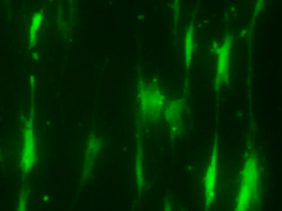
Cell Organization & Morphology
See how nanofiber architecture influences cell morphology, alignment, elongation, and spatial organization across different cell models.
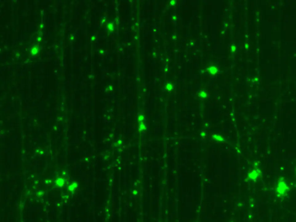
Neuronal Models
Explore how nanofiber density and patterning influence neuronal patterning, alignment, and axonal organization in iPSC-derived human cortical neurons.
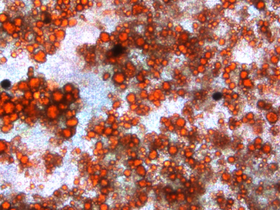
Stem Cell Fate & Differentiation
Explore how nanofiber biophysical cues influence cellular differentiation using existing protocols.
Drug Response
Compare drug response curves from standard 2D plates and USF nanofiber cell culture scaffolds.
Technology
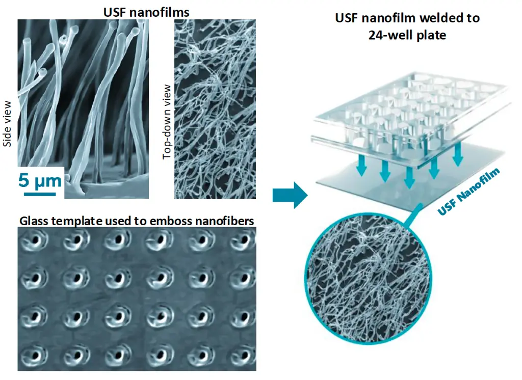
A reusable template or mold is created by drilling billions of cylindrical nanoholes into a glass substrate, placing each hole with 50 nm precision.
The template is used to emboss nanofibers onto a polymer film. The nanofibers form reliably in the template holes, enabling highly precise and customizable fiber spacings and patterns.
Fibers are integrally attached to the film and remain fixed in place.
With further processing, fiber diameter (and therefore stiffness) can be increased from a wispy 50 nm to well over a micron.
This provides the means to customize microenvironments across the entire range of collagen fibrils found in biological scaffolds.
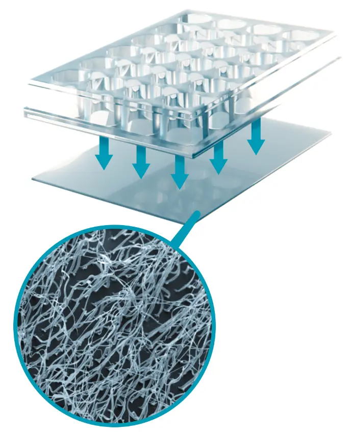
Our 3D nanofiber scaffolds are sealed to the undersides of 2D well plates, which are molded without bottoms but otherwise have standard well plate dimensions.
This allows cell culture to take place in the biomimetic 3D nanofiber microenvironments created by our scaffolds without the need for hydrogels.
These microenvironments are tunable for each application by adjusting the hole patterns and sizes in the template and can be reproduced with high precision.
USF scaffolds are embossed on 125 µm thick polycarbonate cover slips so that cultures can be easily monitored in situ with inverted microscopy.
Other form factors will be available in the future to meet the evolving needs of our customers and the broader marketplace.
USF nanofilms offer inherent advantages over current 3D scaffolds.
Biomimicry
Collagen-like fiber sizes, spacing, density, and orientation create more natural 3D microenvironments for cell culture. The open scaffold structure provides access to cells for nutrients and biochemical signals without requiring hydrogels.
Scalable Production & Versatile Formats
Batch production supports fast prototyping, small form factors, and custom lot sizes. Continuous production offers the potential for larger-scale manufacturing, including rolls wider than 30 cm. The technology can also be adapted to formats beyond well plates, including imaging dishes, T flasks, bioreactors, and parallel-flow devices.
Usability
USF nanofilms work with standard laboratory equipment and protocols and can be functionalized using familiar techniques, thin coatings, and reagents. Transparent well bottoms enable in situ imaging for live-cell observation. Plates can be stored at room temperature.
Tunability & Reproducibility
Precisely controlled fiber spacing, density, and patterns let researchers customize microenvironments for different cell types and applications. Defined architecture can reduce variability associated with the randomness of existing technologies.
Getting Started with Free Tuning Plates
Start with Free Tuning Plates
1. Request your plates.
2. Review preparation guidance and compare conditions using your cells and relevant readouts.
3. Discuss your observations and next configurations with USF.
Discuss Your Application
Have a specific cell model, experimental objective, or customization need? Connect with our team to discuss initial protocols, available configurations, and opportunities to adapt USF nanofilms to your application.
FAQs
Yes. In addition to other intellectual property in the U.S. and abroad, we have broad patent protection for both our foundational technology and its application to biology, including U.S. Patents 11,667,062, 11,155,007, and 10,941,380. Other patents are pending.
Our nanofiber platform is designed to deliver both tunability and reproducibility, with continuous improvements being made and near-term priorities placed on the parameters that most strongly influence biological outcomes. Over time, we expect to provide highly tunable and reproducible control across all key structural parameters and to expand material options to enable application-specific control of properties such as stiffness, wettability, and cell adhesion, in a disciplined and application-driven manner.
We provide highly reproducible control of fiber spacing, density, and pattern, which are tunable by design and reproduced reliably within and across well plates. Fiber spacing can range from below 3 microns (approximately 110,000 fibers/mm²) to arbitrarily large spacings or custom patterns.
Fiber diameter is also highly consistent within a given configuration and from plate to plate. While diameter is not currently offered as a selectable parameter, the diameters used in our standard configurations fall within the central range commonly reported for natural collagen fibrils. Diameter tuning has been demonstrated and may be pursued through focused, application-driven partnerships.
Fiber height is not currently emphasized as a tuning parameter, as most applications are tolerant to variation across the range produced by our standard nanofiber configurations.
Yes. Patterns can be varied from section-to-section or even fiber to fiber. Each fiber can be placed exactly where it is needed to optimize cell behavior for any given application. Corn row patterns are great for aligning neurons, fibroblasts, or other cells.
Yes, we have demonstrated the ability to control fiber stiffness by modifying fiber diameter and material. For now, we are providing PCL fibers in the couple hundred nm range with high reproducibility within and between plates.
Our nanofiber scaffolds are currently made from polycaprolactone (PCL) on optically clear polycarbonate (PC) film substrates. The thickness of the PC is 125 microns, the equivalent of a #0 coverslip. Cells in the well bottoms only contact PCL, which is a bioabsorbable polymer commonly used in FDA-approved in vivo devices. Our well plates are also made from high quality PC. Our imaging dishes are initially made from medical grade polystyrene, but we expect to move to high quality PC over time.
Yes. A wide range of polymers have been used for nanofilm production, and others can be developed for future applications. Our technology is generally compatible with most thermoplastics and some thermosets.
No, hydrogels are unnecessary. Our collagen-like nanofibers already provide a superior 3D microenvironment.
Yes, USF nanofilms are compatible with common coatings, poly-amino acids, and proteins.
Yes. Our well plates are made to standard dimensions and are compatible with liquid handling systems, microplate readers, robotic systems, and other standard lab equipment.
Yes, cells can be viewed in situ through our optically clear well bottoms. The polycarbonate is equivalent to a #0 coverslip specifically for inverted microscopy.
Yes, our nanofibers are integrally and permanently attached to the underlying substrate. They are extremely difficult to remove, even with a razor blade. PCL fibers are bioabsorbable and may eventually degrade in vivo over weeks or months.
Yes, they can be stored at room temperature. While accelerated aging tests have not yet been conducted, we expect them to age similarly to standard 2D well plates.
Yes, other formats can be developed as needed. We will expand our offerings when there is sufficient demand.
Yes, our nanofiber scaffolds can be integrated into various formats, including imaging discs (available now), T flasks, petri dishes, bioreactors, and parallel flow devices. We can batch-produce or scale up with roll-to-roll methods, producing scaffolds in widths greater than 30 cm. We welcome partnerships to develop applications for different needs.
Yes. We are happy to discuss custom solutions, but we will need to prioritize based on complexity and potential impact. If you're interested in customization, contact us to arrange a discussion.
The free set includes three Tuning Plates, each with six nanofiber conditions in a standard 24-well format. Shipping is free within the continental U.S.
Compare how different nanofiber densities and patterns relate to morphology, organization, differentiation, or assay response in your experimental system. Defined, reproducible physical architecture supports controlled comparisons; it does not guarantee reproducible biological results. Outcomes depend on the cell model, preparation, culture conditions, and readouts.
Explore our scientific overview for examples of cell models evaluated with USF nanofiber scaffolds. Evidence is specific to each model and experimental setup. Contact USF to discuss available data for your cell type and the conditions you would like to compare.
A hydrogel is not required to use the nanofiber scaffold. Surface coatings can be used, and the published cell-plating protocol includes a fibronectin coating with options to adapt the coating to the cell model. Choose preparation and coating conditions for your cells and assay; the platform is not a replacement for every hydrogel or organoid workflow.
Visit USF Resources (protocols) for the Cell Plating Protocol and best-practice guidance. Review preparation and coating requirements before starting, and discuss adaptations for your cells and assay with USF.
Review the preparation guidance and plan which conditions, controls, and readouts you will compare. After evaluating the plates with your cells, discuss your observations and possible next configurations with USF. Contact the team with questions about your request or experimental setup.
Our Team

Collin Anderson, CEO
Entrepreneur with 25+ years of experience taking innovations from lab to market. Co-founded, led, and sold a company with a flagship product line he co-invented that generated approximately $300M in lifetime retail sales. Chicago Booth MBA (High Honors) and PhD in Materials Science from Vanderbilt University.

William Hofmeister, Founder, CTO
Inventor of USF’s core technology. Fellow of ASM International, NASA-funded researcher, and International Laser Class Legend. Adjoint Professor of Physics and Astronomy at Vanderbilt University and former Director of the Center for Laser Applications at the University of Tennessee Space Institute.

Clarissa Valdez, Senior Scientist
Neuroscientist with 15+ years of hands-on cell culture and complex disease modeling (iPSC-derived neuronal systems). PhD from Northwestern University; postdoc UT Southwestern. Drives customer data generation and application development.