Nanofiber Scaffolds for Neuronal Cell Culture
Explore how defined nanofiber density and patterning influence neuronal distribution, alignment, and axonal organization in iPSC-derived human cortical neurons.
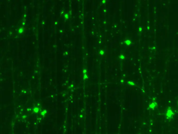
How Nanofiber Architecture Shapes Neuronal Growth
USF nanofilms provide tunable physical architecture for neuronal culture. Compare fiber densities and patterns to explore which conditions suit your model, imaging workflow, and research question.
Neuronal Organization Across Nanofiber Conditions
USF nanofilm technology creates targeted microenvironments that direct and influence neuronal patterning and growth.
Cell model: iPSC-derived human cortical neurons
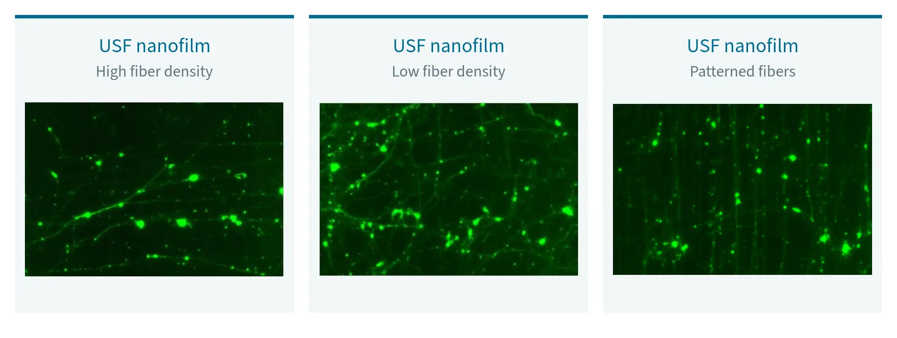
High-Density Nanofiber Spacing: Enhanced isolation of individual neurons and axons. Ideal for imaging and analysis of single cells or axons.
Potential Applications: Neuronal morphology studies, regeneration studies, electrophysiology
Low-Density Nanofiber Spacing: Evenly distributed neuronal cultures similar to traditional 2D culture but with reduced clumping and improved reproducibility and analysis in imaging experiments.
Potential Applications: Multi-cell disease modeling, high-throughput imaging, drug/compound testing, etc.
Patterned Nanofiber Spacing: Corn-row patterned nanofiber spacing directs neurons to grow in aligned, straight lines and provides greater spatial isolation of neurons and axons. This architecture is ideal for axon-focused research.
Potential Applications: Axonal transport and trafficking, live-cell protein dynamics studies, and axonal growth studies.
Screen Six Nanofiber Conditions in One 24-Well Plate
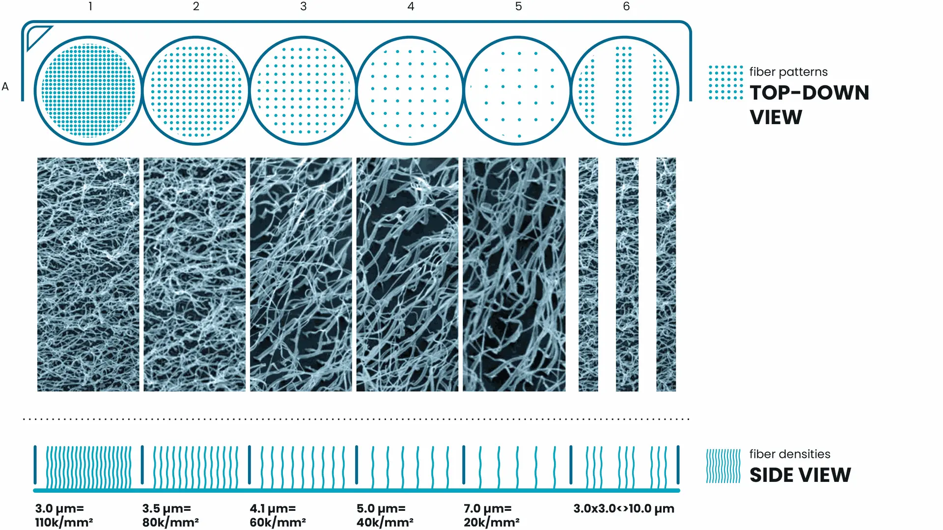
Not every cell model responds to the same nanofiber configuration. USF Tuning Plates provide six defined nanofiber conditions in a familiar 24-well format, helping researchers compare how physical architecture relates to the readouts that matter for their application.
Each of the six columns provides a distinct nanofiber density or pattern. The four rows can be used for different coatings or experimental conditions, allowing you to compare cellular behavior across microenvironments and identify the optimal fiber density for your cells and application.
* Diagram intended for illustrative purposes only.