Nanofiber Scaffolds Influence Cell Organization and Morphology
The extracellular matrix influences how cells spread, align, and organize. Customizable USF Nanofilms recreate that architecture, letting you find the microenvironment that best fits your cells and application.
How Nanofiber Architecture Shapes Cell Organization
Nanofiber architecture provides physical cues that are absent on a flat culture surface. By varying fiber spacing and pattern, USF nanofilms give researchers a controlled way to examine how those cues influence cell behavior.
Primary Cardiomyocytes
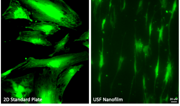
USF nanofilms guide primary neonatal rodent ventricular cardiomyocytes (NRVMs) to self-organize along nanofiber scaffolds, producing in vivo-like alignment and morphology. Cardiomyocytes align directionally along the nanofibers and adopt an elongated morphology resembling in vivo tissue organization.
Cell model: Primary neonatal rodent ventricular cardiomyocytes (NRVMs)
Key observation: Cardiomyocytes showed directional alignment and elongated morphology on the nanofiber scaffold.
USF pilot study, protocol optimization ongoing. Data generated with Cara Barnett and Patrick Oakes, PhD, Loyola University Chicago.
Mouse Embryonic Fibroblasts
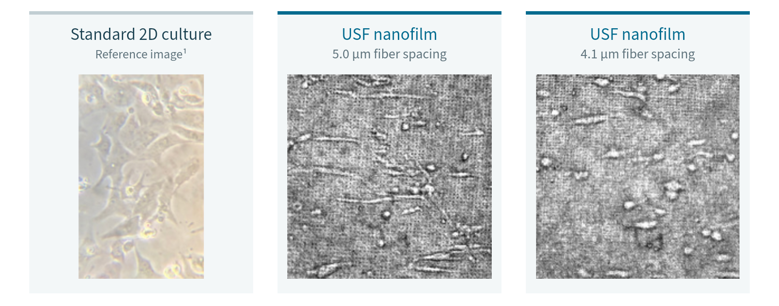
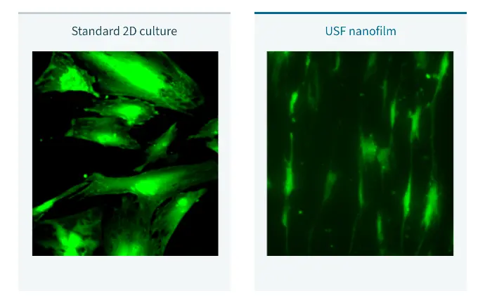
USF nanofilms guide fibroblasts to self-organize along nanofiber scaffolds, producing in vivo-like alignment and morphology that flat 2D surfaces cannot replicate.
Cell model: Mouse embryonic fibroblasts (MEFs)
Key observation: Fibroblasts adopt a spindle-like morphology resembling in vivo tissue organization on USF nanofilm plates.
Screen Six Nanofiber Conditions in One 24-Well Plate
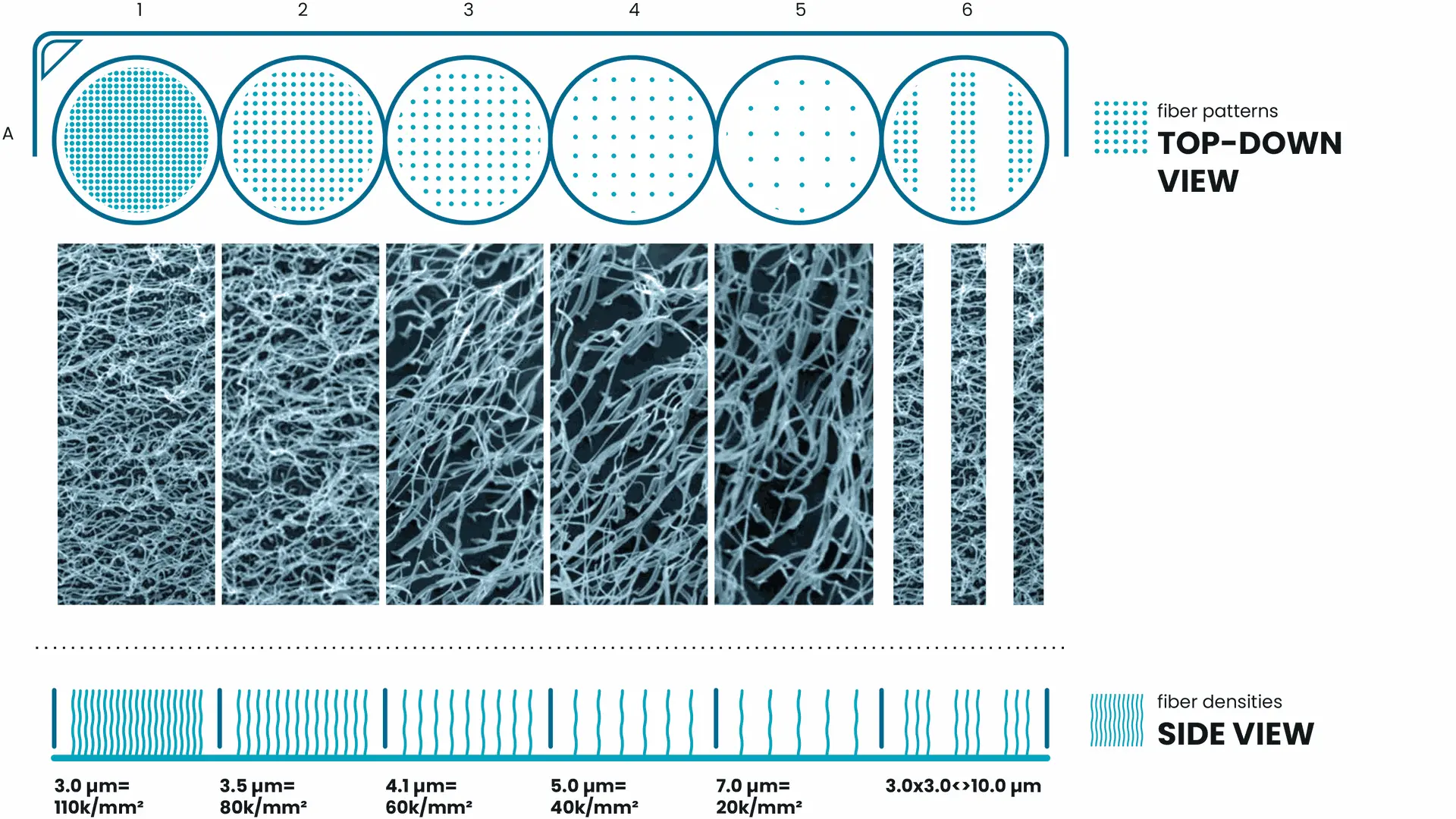
Not every cell model responds to the same nanofiber configuration. USF Tuning Plates provide six defined nanofiber conditions in a familiar 24-well format, helping researchers compare how physical architecture relates to the readouts that matter for their application.
Each of the six columns provides a distinct nanofiber density or pattern. The four rows can be used for different coatings or experimental conditions, allowing you to compare cellular behavior across microenvironments and identify the optimal fiber density for your cells and application.
* Diagram intended for illustrative purposes only.