3D Nanofiber Scaffolds for Drug Response Studies
Compare cell viability and drug sensitivity on standard 2D plates and USF nanofiber scaffolds. Explore HeLa results with doxorubicin and staurosporine.

Does the Microenvironment Influence Drug Response?
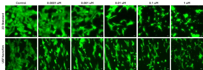
HeLa cells were evaluated across concentration ranges of doxorubicin and staurosporine on standard 2D plates and USF nanofilms. The drug’s target pathway determines whether the microenvironment changes the outcome: doxorubicin showed minimal influence from the microenvironment, while staurosporine showed a strong microenvironment effect.
Ongoing experiments will examine how these results compare to results in 3D organoid
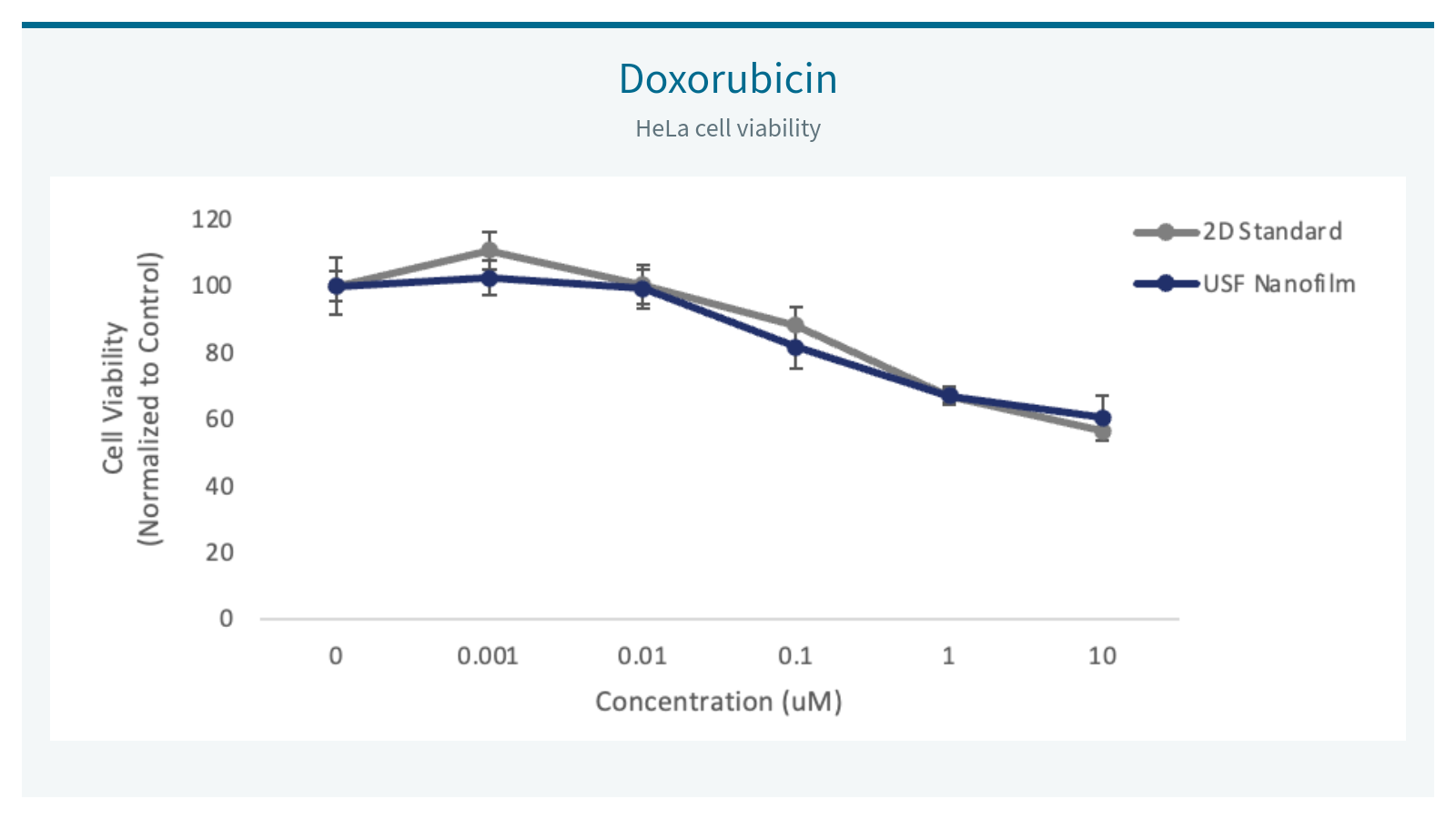
Doxorubicin: Nanofibers Do Not Impede Drug Activity or Delivery
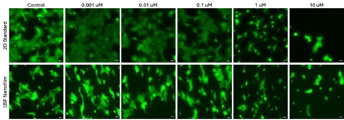
Doxorubicin produced similar viability curves on standard 2D plates and USF nanofilms. Doxorubicin acts directly on DNA, making it less dependent on cell shape, adhesion, or microenvironment. Consistent potency across both microenvironments suggests that nanofibers do not impede drug activity or delivery.
Cell model: HeLa cells
Key observation: Doxorubicin response was similar across standard 2D and USF nanofilm conditions.
Staurosporine: Strong Microenvironment Effect
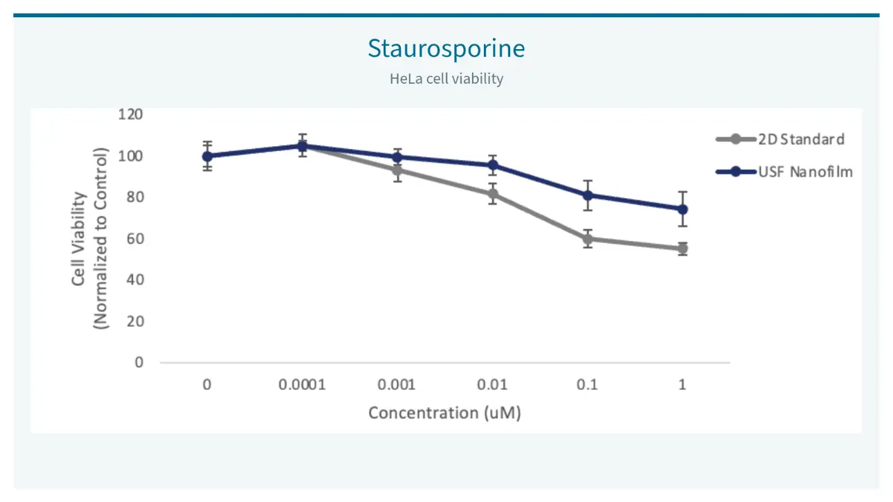
Staurosporine produced clear, sustained separation of viability curves on standard 2D plates and USF nanofilms, showing a meaningful decrease in sensitivity to treatment on USF nanofilms.
Staurosporine is a broad protein kinase inhibitor and strong apoptosis inducer that blocks survival-signaling pathways. These pathways are strongly influenced by cell–matrix interactions and cytoskeletal organization. USF nanofilms appear to capture effects of the extracellular matrix (ECM) on drug response typically missed by standard plates.
Cell model: HeLa cells
Key observation: Lower sensitivity to staurosporine was observed on USF nanofilms under the tested conditions. USF nanofilms appear to capture effects of ECM on drug response typically missed by standard plates.
Screen Six Nanofiber Conditions in One 24-Well Plate
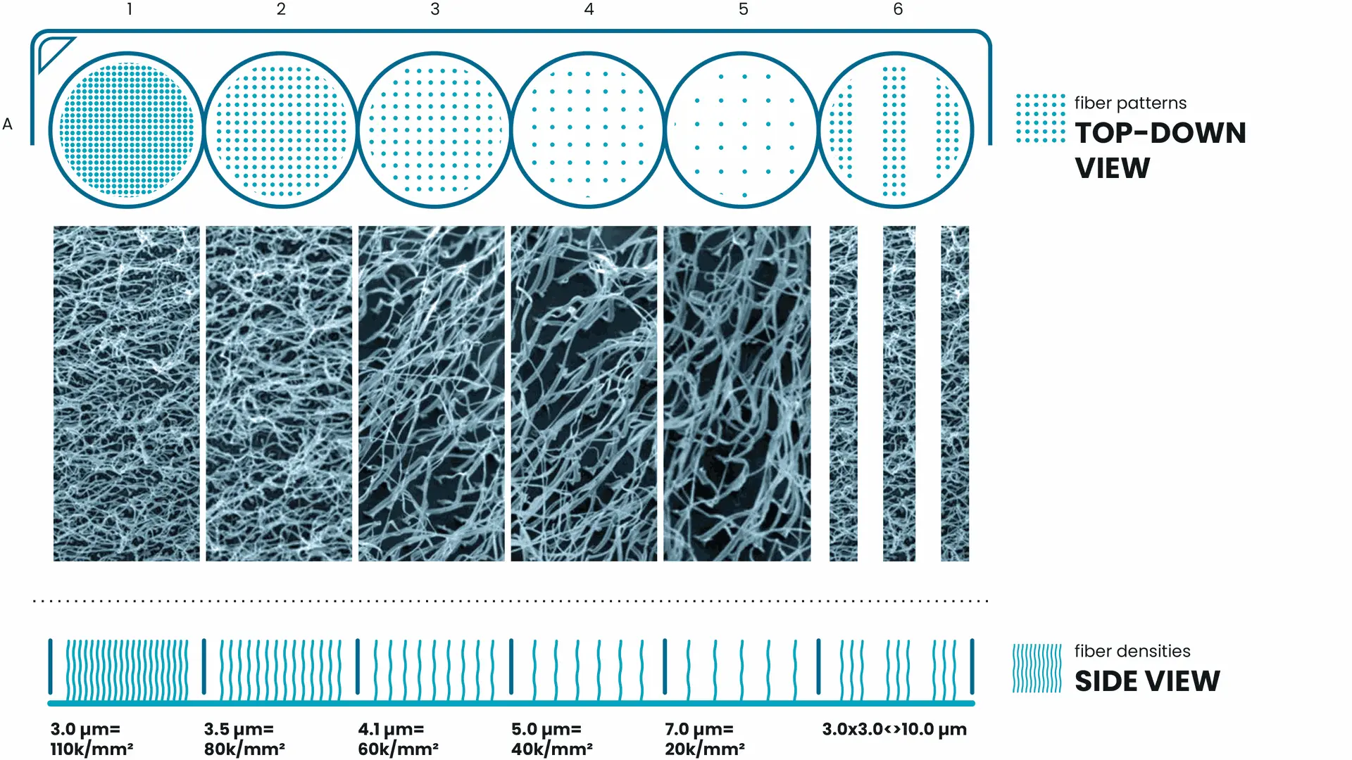
Not every cell model responds to the same nanofiber configuration. USF Tuning Plates provide six defined nanofiber conditions in a familiar 24-well format, helping researchers compare how physical architecture relates to the readouts that matter for their application.
Each of the six columns provides a distinct nanofiber density or pattern. The four rows can be used for different coatings or experimental conditions, allowing you to compare cellular behavior across microenvironments and identify the optimal fiber density for your cells and application.
* Diagram intended for illustrative purposes only.