Nanofiber Scaffolds for Stem Cell Fate and Differentiation
USF nanofilm scaffolds introduce tunable physical cues into existing differentiation protocols, helping researchers explore how the microenvironment influences stem cell fate and differentiation.
How Physical Microenvironments Influence Stem Cell Fate
Cell phenotype and differentiation are influenced by both biochemical signals and the surrounding physical microenvironment. Explore early observations from adipogenic and neuronal cell models cultured on USF nanofilms.
Adipogenic Differentiation
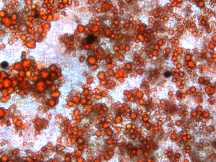
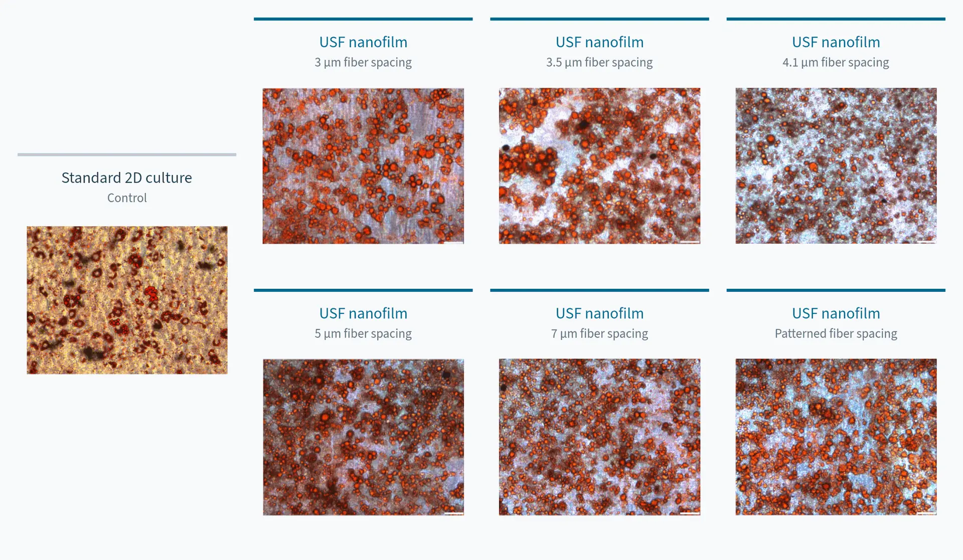
USF nanofilm plates enhance adipose cell differentiation. Adipose cells differentiated from 3T3 cells showed visibly greater lipid accumulation across all six USF nanofilm conditions than on standard 2D plates, with all other protocol conditions held constant. USF nanofilm biophysical cues work synergistically with the existing biochemistry to promote more effective differentiation. Quantification is in progress.
Cell model: Adipose cells differentiated from 3T3 cells
Key observation: Visibly greater lipid accumulation across all six USF conditions.
Pilot experiment; protocol optimization and quantification are ongoing. Data generated with Angelica Ross and Marcella Vaicik, PhD, Illinois Institute of Technology.
Neuronal Differentiation
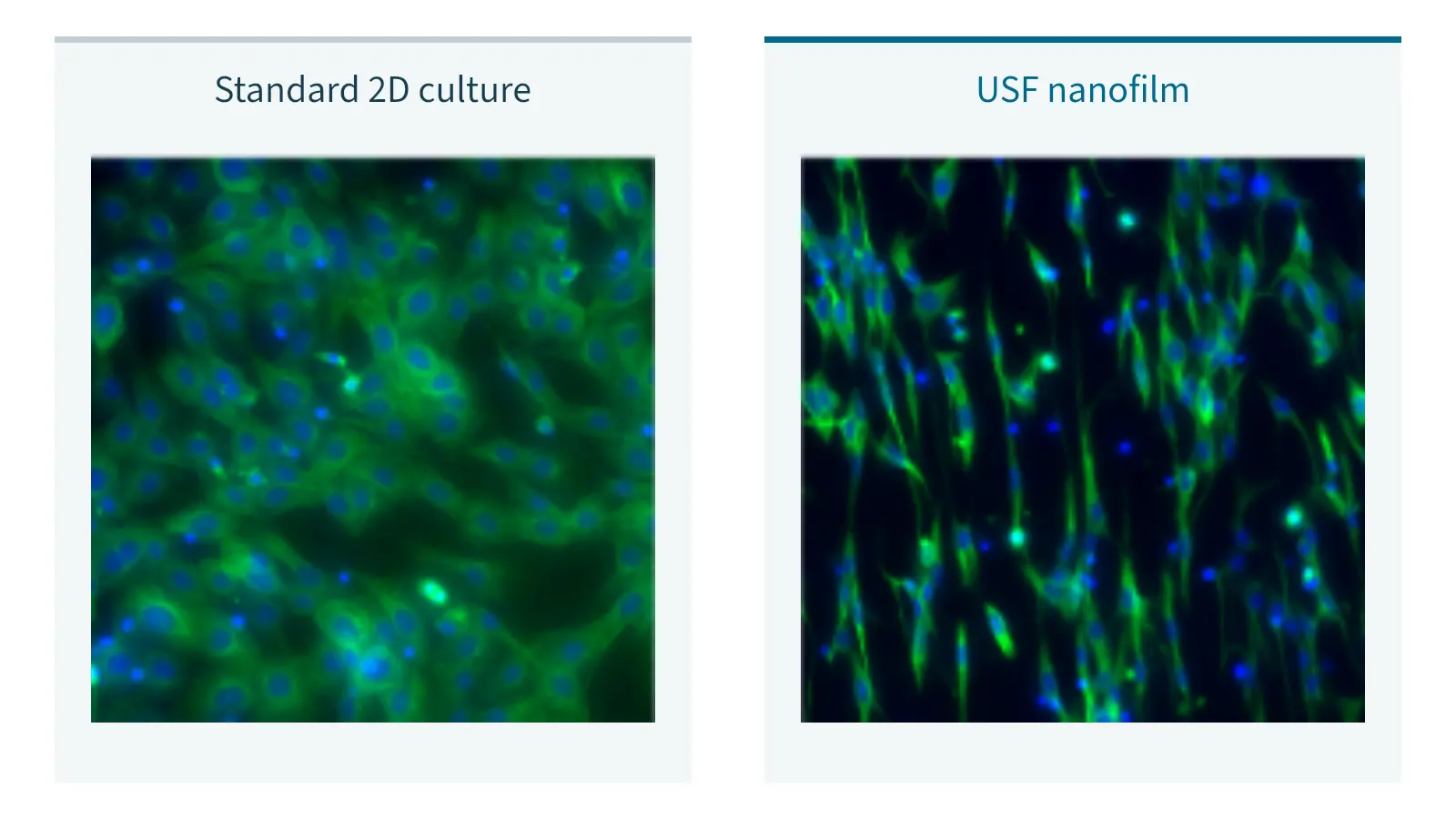
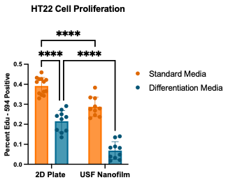
USF nanofilm plates enhance HT22 cell differentiation toward a more neuronal phenotype. Biochemical differentiation signals combined with USF nanofilm biophysical cues work synergistically to promote more effective differentiation than on standard 2D plates. HT22 cells showed decreased proliferation, a hallmark of neuronal differentiation.
Cells on USF nanofilms also adopted neuronal-like morphology.
Cell model: HT22 cells
Key observation: Decreased proliferation and neuronal-like morphology under the tested differentiation protocol.
Screen Six Nanofiber Conditions in One 24-Well Plate
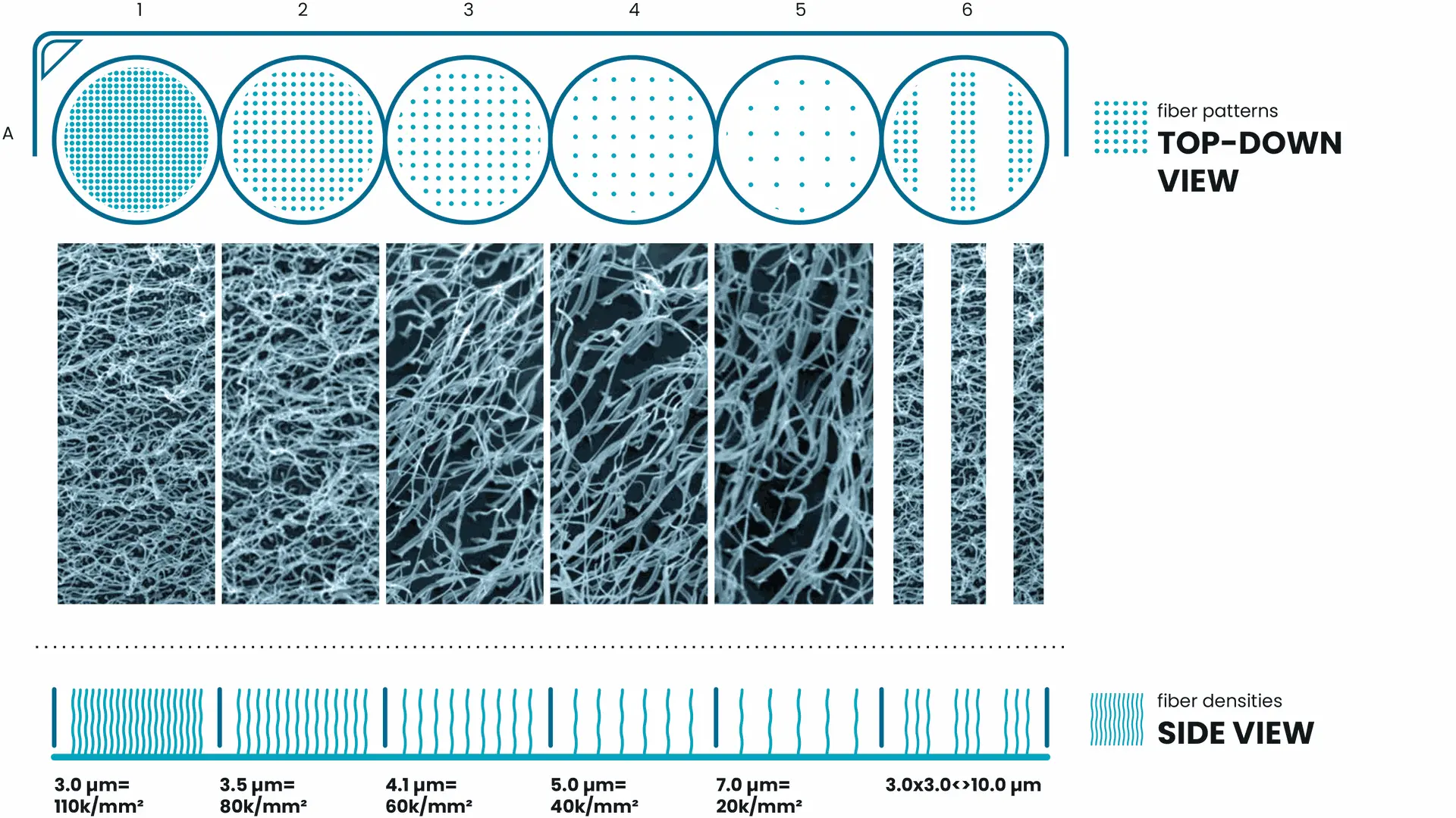
Not every cell model responds to the same nanofiber configuration. USF Tuning Plates provide six defined nanofiber conditions in a familiar 24-well format, helping researchers compare how physical architecture relates to the readouts that matter for their application.
Each of the six columns provides a distinct nanofiber density or pattern. The four rows can be used for different coatings or experimental conditions, allowing you to compare cellular behavior across microenvironments and identify the optimal fiber density for your cells and application.
* Diagram intended for illustrative purposes only.